This page provides information on the types of tests and investigations that may be required leading to a diagnosis of MUO/CUP. These will vary depending on the symptoms and the results of other tests. This section also explains the role of a pathologist and the broader role of genomics in helping diagnose CUP or a primary cancer.

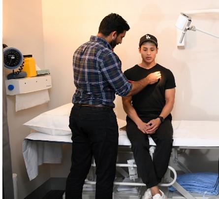
Your doctor will conduct a physical examination of your body. Your doctor will pay particular attention to the areas of your body where you are experiencing symptoms, which will be guiding the exam. This can include looking for lumps on your body, feeling for swollen lymph nodes or anything that might be unusual.
Your doctor will also have a discussion with you about your health history and your family history. This can include:

A biopsy is a procedure to obtain a small amount of tissue for testing, with the overall purpose of identifying the type of cancer and, if possible, the original location of the cancer. This may help to guide your doctors in making a diagnosis and choosing the best treatment option. However, if the cancer is too hard to reach or if you are too unwell for the procedure, your doctor might decide against performing a biopsy.
There are multiple ways to obtain a tissue sample and the method used will depend on the type of mass and location in the body. Depending on the method, you will have local anaesthesia to numb the area or general anaesthesia to make you unconscious during the procedure.
Role of the Pathologist in diagnosing CUP
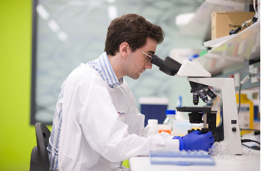
After you have a biopsy, it will be sent to a laboratory for testing. The sample of tissue will be looked at under a microscope by a specialist doctor called a pathologist. A pathologist is a doctor specialising in the diagnosis of disease based on examination of tissues and fluids removed from the body. Upon examination, the pathologist determines if the tissue sample contains normal, pre-cancerous or cancerous cells and then writes a report on their findings (pathology report).
The pathologist will try and identify the type of cancer present in the biopsy, determine the original location of the cancer, and provide information that may help to identify the best treatment option. This helps to guide the clinical team in making a diagnosis and choosing the best treatment. Cancer changes the appearance of healthy cells, making them look abnormal. Pathologists can also identify which cells do not belong in the tissue or organ that it has spread to and are able to identify it as a secondary cancer. However, in MUO or CUP, the primary cancer is not easily recognisable. These cells may have become so abnormal that it is difficult to identify where they came from. These cells are called poorly differentiated or undifferentiated.
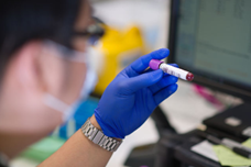
Further testing may be required, such as immunohistochemistry, where the pathologist uses special stains to locate specific proteins that are linked to known cancer types (e.g., adenocarcinoma, squamous cell carcinoma).
Watch and listen to a medical pathologist share her expert insights about the role of a pathologist in diagnosing CUP.

Depending on your situation, your doctor might recommend additional scans. You will need to inform the doctor before having any scans if you have any allergies or past reactions, chronic conditions such as diabetes, or are pregnant or breastfeeding.
X-ray
| How it works | X-rays deliver low-energy beams of radiation to create images of parts of the body to produce the pictures. X-ray beams are absorbed differently by structures in the body, such as bones that absorb radiation easily (appearing white in the picture) or soft tissues like organs that don’t absorb radiation as easily and will appear gray. |
|---|---|
| How long | 10-30 minutes. |
| What happens | Some x-rays use dye (contrast) to improve the image and make certain structures easier to see. Depending on what part of the body is being x-rayed, you might be asked to lie, sit or stand. You will hold still in front of or on a machine while the images are taken. |
| Special notes | The procedure is painless with only a small dose of radiation. The contrast dye is generally very safe and reactions are uncommon; however, people with diabetes and kidney disease should inform their doctor as they will have a slightly greater risk of reaction. |
Mammogram
| How it works | An x-ray machine that is designed only for mammograms will use low-dose x-ray beams to create an image of the inside of the breast. |
|---|---|
| How long | 10-30 minutes. |
| What happens | You will stand in front of an x-ray machine that is specially designed for mammograms. Your breast is placed between two x-ray plates, one at a time, which will press together firmly to spread the tissue so a clear image can be taken. This also prevents movement so the image does not become blurred. Usually two different views are taken of each breast. During the brief period when you are exposed to the x-ray beams, you will be asked to stand still and hold your breath. |
| Special notes | The procedure can be uncomfortable when the x-ray plates are pressed together. If the discomfort becomes too much, notify the technologist (the person performing the procedure). You should notify your doctor if you are pregnant (or believe you are) or breastfeeding. |
Ultrasound
| How it works | An ultrasound is a generally non-invasive imaging test that uses soundwaves to create a picture of the inside of the body. These soundwaves echo when they meet something solid, such as an organ or tumour and a computer will create the image. |
|---|---|
| How long | 10-20 minutes. |
| What happens | A gel is spread on your skin in the required area and a handheld device called a transducer will pass across the surface. The transducer produces high-frequency sound waves which pass through the gel and into your body. The soundwaves bounce off structures in the body and back to the transducer, converting them into an image. Some transducers are wands that can be inserted into a body cavity. |
| Special notes | The procedure is usually painless, but it can be uncomfortable when a transducer is inserted into a body cavity. The soundwaves produced during an ultrasound will not be able to be heard. |
CT scan (computerised tomography scan)
| How it works | A CT scan uses x-ray beams and a computer to create detailed pictures of the inside of the body. The scanner is large and shaped like a doughnut. CT scans will provide more detail than an x-ray as it takes dozens to hundreds of images of your body. This provides a clearer image of soft tissues in the body, which don’t always show up on a regular x-ray, and also shows organs and other structures that overlap or are hidden. |
|---|---|
| How long | 30-60 minutes (mostly due to preparation). |
| What happens | Before the scan, you may be given a drink or injected with a dye (contrast) to highlight certain structures in your body. This will make the images clearer. You will usually lie still on a table that moves into the scanner. It is important to lie still as movement could cause blurry or less clear images. |
| Special notes | The procedure is painless; however, the dye can make you feel hot or flushed and leave a bitter taste in your mouth. There is a small risk of having an allergy-like reaction to the dye, such as a rash, itchiness, nausea, sweating or difficulty breathing. If you know that you have an allergy to the dye or contrast, or if you are diabetic or have some known kidney issue, inform your doctor. You should also notify your doctor if you are pregnant (or think you may be) as CT scans use slightly higher levels of radiation than x-rays. |
PET scan (positron emission tomography scan)
| How it works | PET scans use a low-dose radioactive solution to measure cell activity in different parts of the body. The one most commonly used is fluorodeoxyglucose (FDG) which is a simple sugar (glucose) that has been made radioactive. This means that it will give off energy in the body to be seen by the scanner. Cancer cells grow quickly, and as such, need a lot of energy (or a lot of sugar). The increased amount of sugar needed in these areas will show up as “hot spots”. |
|---|---|
| How long | About 2 hours. |
| What happens | You will be asked to remove anything metal from your body as it interferes with the images of the scanner. You are injected in the arm with a small amount of the radioactive solution and wait 30-90 minutes to allow the solution to spread through your body. You will be lying down, usually on your back, on a couch while it moves backwards and forwards in the scanner. The scanner is large and doughnut shaped. As you move through the scanner, it will take pictures of your body. |
| Special notes | You need to notify your doctor if you are pregnant or have any possibility of being so or are breastfeeding. The specialist will also tell you to avoid children and pregnant women for a few hours after the scan. It is very rare to have an allergic reaction to the radioactive solution. The solution will leave your body in your urine within a few hours. |
PET-CT Scan
| How it works | This test combines both a PET scan and a CT scan and will provide more detailed information about the cancer. See above for information about PET scans and CT scans. |
|---|---|
| How long | About 2 hours. |
Bone scan
| How it works | A bone scan will look for changes in your bones, using a radioactive tracer (dye) injected into your bloodstream. It can look at one particular joint or bone, but it is more usual for the entire body to be scanned for cancer patients. |
|---|---|
| How long | The test takes between 30-60 minutes, but you will be required to be at the hospital for several hours. |
| What happens | You are injected in the arm with a small amount of radioactive dye. You will then wait 2–3 hours for it to move through your bloodstream and collect in your bones. The dye tends to collect more in areas where the bone is breaking down or repairing itself and will appear darker on the scan than the other bones. You will lie down on a couch and must keep very still while going through the scanner. A large camera connected to an arm-like device will scan you and pick up on the radioactivity. |
| Special notes | If you are pregnant or breastfeeding, notify your doctor and the specialist conducting the scan. The dye leaves your body through your urine after a few hours and you will be told to avoid children and pregnant women for a number of hours. |
MRI scan (magnetic resonance imaging scan)
| How it works | An MRI scan uses a magnet and radio waves to take detailed pictures of a particular area inside your body. |
|---|---|
| How long | 30-90 minutes. |
| What happens | You may be injected with a dye (contrast) depending on the scan you are getting to help the tissues show up clearer on the image. You will lie down on a table and be given earplugs or headphones as the MRI scanner is very noisy. You will also be given a buzzer so you can talk to the specialist who is in a different room whenever you press the button. The scanner is a narrow metal cylinder that is open at both ends. During the scan, the table will move in and out of the tunnel. Depending on the scan, you might have leads (wires or stickers) placed on your chest to monitor your heartbeat, a plastic tube attached to your finger to monitor your breathing, or a needle inserted into a vein if any medicine is needed. The part of the body that is being scanned will be gently secured so that the images collected are clear. |
| Special notes | If you might be pregnant, have kidney problems or do not like closed spaces, you should talk to your doctor. If you feel anxious in closed spaces, the doctor may give you some medicine to keep you calm. If there are any metallic objects in your body, such as a pacemaker, you will not be able to have an MRI – notify your doctor. An allergic reaction to the dye (contrast) is very rare, but any reactions are usually mild. |
[Adapted from Cancer Council Australia. Understanding Cancer of Unknown Primary. IVE Group; 2022]
Further information about about Medical Procedures:

| Tumour Marker | Types of Cancer | |
|---|---|---|
| Prostate-specific antigen | PSA | Prostate cancer |
| Alpha-fetoprotein | AFP | Testicular and liver cancers |
| Human chorionic gonadotropin | HCG | Testicular cancer and a rare type of ovarian cancer |
| Carcinoembryonic antigen | CEA | Bowel, lung, pancreatic, stomach, ovarian, breast, thyroid, and liver cancers |
| Cancer antigen 125 | CA125 | Ovarian, endometrial, fallopian tube, and peritoneal cancers |
| Cancer antigen 19-9 | CA 19-9 | Pancreatic, stomach, bile duct, gallbladder, and ovarian cancers |
| Cancer antigen 15-3 | CA 15-3 | Breast cancer |
Your doctor will only recommend additional tests (such as an endoscopy) if they are appropriate and may help locate the primary cancer. This will be based on your individual situation, such as your presenting symptoms and results of previous tests.
Endoscopy
An endoscopy is a procedure that uses an endoscope (a thin, flexible tube with a light and camera on the end) to look inside the body for any abnormalities. The endoscope is inserted through a natural opening (such as the mouth) or through a small cut made by a surgeon. The endoscope can also be used to take a biopsy if your doctor notices anything suspicious. The most common endoscopies performed for CUP include gastroscopy and colonoscopy.
| Endoscopy Type | What is checked |
|---|---|
| Bronchoscopy or endobronchial ultrasound (EBUS) | Lungs or respiratory tract (airways) |
| Colonoscopy | Colon (large bowel) |
| Cystoscopy | Bladder |
| Gastroscopy | Oesophagus, stomach and first part of the small bowel |
| Hysteroscopy | Vagina and cervix |
| Laparoscopy | Abdominal cavity, liver, bowel, uterus and ovaries |
| Laryngoscopy | Larynx (voice box) |
| Sigmoidoscopy | Lower part of the colon (large bowel) |
| Thoracoscopy | Lungs |
The Role of Genomic Testing in CUP
Watch and listen to genomic experts share their insights about genomic testing for CUP patients.

Here are the links to other information pages to learn more about different aspects of diagnosis. You may also use the quick links on the right side of the page to navigate.

Most people are diagnosed with cancer of unknown primary (CUP) after they have symptoms or become unwell. Some people may be diagnosed during tests for another health condition. When cancer is suspected, you might be referred for tests or to a specialist.

The treatment you have depends on a number of things, including where the cancer is and your general health. A team of doctors and other professionals discuss the best treatment and care for you. The main treatment for Cancer of Unknown Primary is cancer drugs, most commonly chemotherapy. You may also have radiotherapy to help to control your symptoms and hormone therapy.
